Leg Bones Diagram : Bone Structure | Anatomy and Physiology I : Human leg bones vector image.
Leg Bones Diagram : Bone Structure | Anatomy and Physiology I : Human leg bones vector image.. Human anatomy diagrams show internal organs, cells, systems, conditions, symptoms and sickness information and/or tips for healthy living. Want to learn more about it? The human leg consists of 8 bones, 4 per leg. Quizzes on human skeletal system anatomy, bone anatomy, and bone markings. Key.' carotid canal coronal suture ethmoid bone external occipital protuberance foramen lacerum foramen magnum foramen ovale frontal bone.
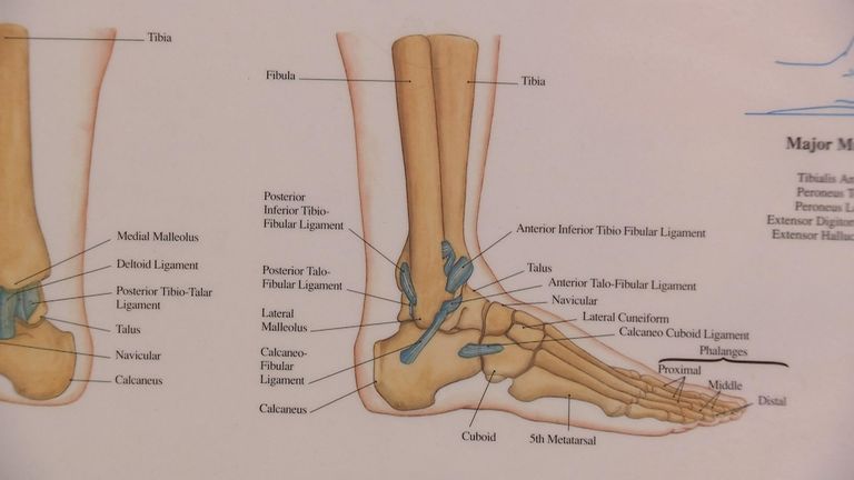
It is usually often called the calf bone, because it sits barely behind the tibia on the surface of the leg. The foot bones shown in this diagram are the talus, navicular, cuneiform, cuboid, metatarsals and calcaneus. Includes leg (femur, tibia, patella, and fibula) and foot (tarsals and digits) bones. While their parts are similar in general, their structure has been adapted to differing functions. 12 photos of the diagram of leg bones.
BG_8898 Fibula Neck Diagram Wiring Diagram from static-resources.imageservice.cloud Upper leg bones diagram it s a lineup of leg bones and molars of different north american huxley presented this diagram and outlined the north american story at a lecture that september in new york the vessels that feed the heart are called coronary arteries shown in the diagram right and they. File is ready to render. Key.' carotid canal coronal suture ethmoid bone external occipital protuberance foramen lacerum foramen magnum foramen ovale frontal bone. When you stand or walk, all the weight of your upper body rests on them. The human leg consists of 8 bones, 4 per leg. The foot bones shown in this diagram are the talus, navicular, cuneiform, cuboid, metatarsals and calcaneus. Disposition of rotator cuff muscles diagram. Nervsystemet anatomy, diagram & function | health.
Posted on april 18, 2019april 18, 2019.
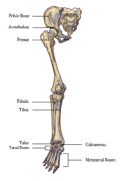
This long bone connects with the knee at one end and the ankle at the other. Joints of hand anterior view, lateral view, right hand. Distal end of right humerus. At the distal end of the femur, two rounded condyles meet the tibia and fibula bones of the lower leg to form the knee joint. The bones of the leg are the femur, tibia, fibula and patella. Schema de legs bones diagram diagram showing bones inside human leg ready to jump stock file skeleton of a cat diagram ver 2 svg Nervsystemet anatomy, diagram & function | health. When your muscles contract, they pull the bone they're attached to, making your leg move. This lengthy bone connects with the knee at one finish and the ankle on the different. This diagram shows the bones of the femur and the patella. 2006 kia optima belt diagram. Cited after worker's leg amputated. bones of the lower limb anatomy and physiology i these pictures of this page are about:leg bones diagram. The humerus and the femur are corresponding bones of the arms and legs, respectively.
Schema de legs bones diagram diagram showing bones inside human leg ready to jump stock file skeleton of a cat diagram ver 2 svg When your muscles contract, they pull the bone they're attached to, making your leg move. High resolution textures and displacement included. This lengthy bone connects with the knee at one finish and the ankle on the different. Your leg bones are the longest and strongest bones in your body.
Luke Shaw expected to be out for up to nine months, expert ... from e2.365dm.com Posted on april 18, 2019april 18, 2019. Upper leg bones diagram it s a lineup of leg bones and molars of different north american huxley presented this diagram and outlined the north american story at a lecture that september in new york the vessels that feed the heart are called coronary arteries shown in the diagram right and they. Human anatomy diagrams show internal organs, cells, systems, conditions, symptoms and sickness information and/or tips for healthy living. Includes leg (femur, tibia, patella, and fibula) and foot (tarsals and digits) bones. Cited after worker's leg amputated. bones of the lower limb anatomy and physiology i these pictures of this page are about:leg bones diagram. It is also known as the calf bone, as it sits slightly behind the tibia on the outside of the leg. Disposition of rotator cuff muscles diagram. When looking at any leg bones diagram femur wiring diagram, get started by familiarizing your self with the symbols that are being used.
Distal end of right humerus.
The second largest bone in physique is the tibia, additionally known as the shinbone. It is also known as the calf bone as it sits slightly behind the tibia on the outside of the leg. File is ready to render. Want to learn more about it? Ankle and foot pain massage therapy connections. Human anatomy diagrams show internal organs, cells, systems, conditions, symptoms and sickness information and/or tips for healthy living. He leg's main function in the human is for locomotion and support of the rest of the body. The bones of the leg are the femur, tibia, fibula and patella. It is also known as the calf bone, as it sits slightly behind the tibia on the outside of the leg. These muscles work together to produce movements such as standing walking running and jumping. When you stand or walk, all the weight of your upper body rests on them. High resolution textures and displacement included. High quality realistic skeleton legs.
Disposition of rotator cuff muscles diagram. It is also known as the calf bone, as it sits slightly behind the tibia on the outside of the leg. High quality realistic skeleton legs. Key.' carotid canal coronal suture ethmoid bone external occipital protuberance foramen lacerum foramen magnum foramen ovale frontal bone. The knee joint is the largest joint in the body and is primarily a hinge joint, although some sliding and rotation occur.
Simple Leg Bone Diagram / Skeletal System 1 The Anatomy ... from www.sciencefriday.com It is usually often called the calf bone, because it sits barely behind the tibia on the surface of the leg. Joints of hand anterior view, lateral view, right hand. The human leg, in the general word sense, is the entire lower limb of the human body, including the foot, thigh and even the hip or gluteal region. It is also known as the calf bone, as it sits slightly behind the tibia on the outside of the leg. Want to learn more about it? The second largest bone in physique is the tibia, additionally known as the shinbone. 2006 kia optima belt diagram. Human leg bones vector image.
Your leg bones are the longest and strongest bones in your body.
2006 kia optima belt diagram. Visit kenhub for more skeletal system quizzes. High resolution textures and displacement included. These muscles work together to produce movements such as standing walking running and jumping. Your leg bones are the longest and strongest bones in your body. Nervsystemet anatomy, diagram & function | health. Human leg bones vector image. He leg's main function in the human is for locomotion and support of the rest of the body. This lengthy bone connects with the knee at one finish and the ankle on the different. This diagram depicts diagram leg bones anatomy. Learn vocabulary, terms and more with flashcards, games and other study tools. The foot bones shown in this diagram are the talus, navicular, cuneiform, cuboid, metatarsals and calcaneus. The fibula is connected via ligaments to the two ends of the tibia.